

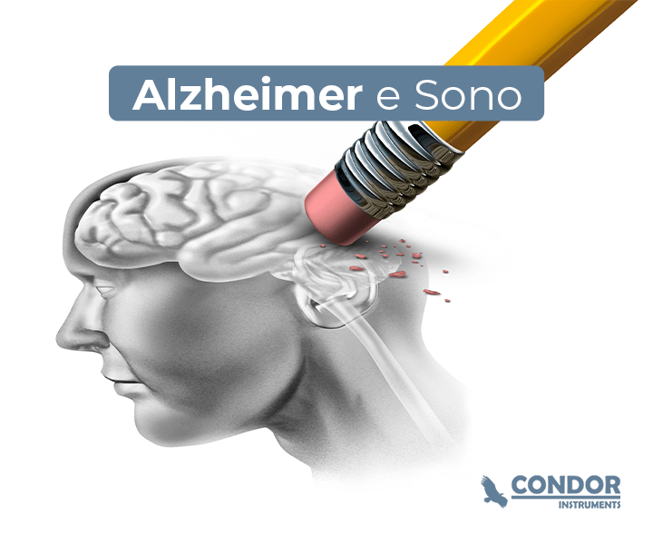
Alzheimer’s disease is a neurodegenerative disease that was first documented in 1906 by Alois Alzheimer.SORIA LOPEZ; GONZALEZ; LEGER, 2019Degeneration occurs mainly in areas responsible for language, memory, visual processing, and executive functions.KNOPMAN; AMIEVA; PETERSEN; CHÉTELAT et al., 2021Diagnosis is complicated by the late onset of observable symptoms, which occur between 10 to 20 years after the onset of degeneration.SPERLING; AISEN; BECKETT; BENNETT et al., 2011and the greatest risk factor for the development of the disease is age. There are still controversies regarding the causes of the disease; however, the most prevalent hypothesis is that the disease occurs from the accumulation of beta-amyloid protein in the brain (SELKOE, 2004Although Alzheimer’s disease is the leading cause of dementia, this condition can be triggered by other neurodegenerative pathologies, especially in the elderly (KAPASI, DECARLI, SCHNEIDER, 2017Furthermore, the prevalence of dementia is high and tends to rise further, from 50 million people in 2010 to 113 million people worldwide in 2050.BRODATY; BRETELER; DEKOSKY; DORENLOT et al., 2011Therefore, considering the debilitating nature of the disease and its high prevalence, the objective of this article is to elucidate the relationships between Alzheimer’s disease and the sleep-wake cycle.
Sleep and wake-sleep rhythm disorders are common in patients with Alzheimer’s disease. These patients exhibit imbalances regarding the onset time and duration of sleep, which manifest as increased sleep latency and awake time during the night, as well as sleep episodes during the daytime, which can eventually lead to a phase inversion of sleep in these patients. Analyzing sleep architecture, it is observed that these patients show a reduction in the proportion of REM sleep compared to age-matched control individuals, leading to chronic REM sleep restriction.PETIT; GAGNON; FANTINI; FERINI-STRAMBI et al., 2004Not only that, but these patients also show a reduced proportion of slow-wave sleepMARTIN; LOEWENSTEIN; KAYE; EBERT et al., 1986).
These changes in temporal organization and sleep structure occur early in the course of the disease. There are studies in rats demonstrating that beta-amyloid protein deposition leads to changes in sleep architecture, sleep phase delay, altered nocturnal activity levels, and reduced REM sleep. These effects were, in some cases, reversed after beta-amyloid immunotherapy, demonstrating the role this protein plays in patients.ROH; HUANG; BERO; KASTEN et al., 2012).

These disorders are worsened by age, especially if these individuals have other clinical conditions. This leads them to reduce physical activity levels, have irregular meal times, and consequently, generate few stimuli for synchronizing the wake-sleep rhythm. Reinforcing this component, institutionalized patients often have low light exposure, and finally, medications for depression, hypertension, and heart disease can lead to wake-sleep rhythm disorders.JU; LUCEY; HOLTZMAN, 2014These factors amplify signals that negatively contribute to sleep quality and wake-sleep rhythm stability.
On the other hand, there are studies demonstrating that sleep restriction and/or poor sleep quality are predisposing factors for the development of cognitive disordersFAUBEL; LOPEZ-GARCIA; GUALLAR-CASTILLON; GRACIANI et al., 2009A prospective study evaluated the sleep of volunteers using actigraphy, and the results showed that sleep fragmentation increased the risk for the development of Alzheimer’s disease (STERNICZUK, THEOU, RUSAK, ROCKWOOD, 2013In both rats and humans, soluble beta-amyloid levels fluctuate throughout wakefulness-sleep, with levels increasing during wakefulness and decreasing during sleep. Thus, acute sleep deprivation increases beta-amyloid concentration, and moreover, chronic sleep restriction accelerates the deposition of beta-amyloid into insoluble plaques.ANCOLI-ISRAEL; COLE; ALESSI; CHAMBERS et al., 2003).
Conclusion
Alzheimer’s disease is an extremely debilitating condition for the individual, and with regard to sleep, evidence indicates that nocturnal insomnia, nighttime agitation, and excessive daytime sleepiness affect 25 to 40% of patients with Alzheimer’s disease, such that the severity of these disturbances correlates with the severity of the disease. As previously mentioned, circadian rhythms exhibit a reduction in the amplitude of endogenous rhythms and a phase delay in these rhythms, especially in advanced stages of the disease. Sleep disturbances occur early in the course of the disease, a fact that contributes to the progression of the pathology. Thus, it is important that these patients be monitored and receive specific care, especially regarding the assessment of biological rhythms and sleep disturbances.
How to assess the sleep of these patients
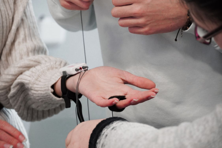
Polysomnography is considered the gold standard for sleep evaluation, however, its use in patients with Alzheimer’s disease is generally difficult because it requires patient cooperation, and in many cases, especially in advanced stages of the disease, patients cannot tolerate laboratory procedures. Similarly, the application of questionnaires, such as the Pittsburgh Sleep Quality Index (Pittsburgh Sleep Quality Index – PSQI have little practical value, as these patients tend to underestimate existing sleep and rhythm disturbances, even in the early stages of the diseaseMOST; ABOUDAN; SCHELTENS; VAN SOMEREN, 2012leading to biased results.
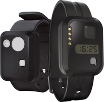
Thus, actigraphy becomes a more suitable procedure for evaluating sleep and the wake-sleep rhythm in these patients, as it is a less invasive technique. Actigraphy is a technique that can be used to evaluate sleep patterns and the wake-sleep rhythm. The actigraph is a device similar to a wrist watch that contains light, motion, and temperature sensors. Therefore, through this information, it is possible to extract data regarding variables such as total sleep time, sleep onset latency, wake time after sleep onset, sleep efficiency, wake time, and rhythm-related variables such as cosinor, spectrogram, periodogram, and non-parametric variables like L5, M10, IS, and VI.